Sunday, 29 January 2017

Pagets Disease

Paget's Disease:

Paget’s disease of bone is a condition characterized by abnormal and anarchic resorption and deposition of bone, resulting in distortion and weakening of the affected bones

LINK: PAGET'S DISEASE PPT

Osteogenesis Imperfecta

Definition:

Osteogenesis imperfecta (OI) is a genetic disorder characterized by bones that break easily, often from little or no apparent cause. A classification system of different types of OI is commonly used to help describe how severely a person with OI is affected. For example, a person may have just a few or as many as several hundred fractures in a lifetime.

Prevalence:

While the number of people affected with OI in the United States is unknown, the best estimate suggests a minimum of 20,000 and possibly as many as 50,000.

Diagnosis:

OI is caused by genetic defects that affect the body’s ability to make strong bones. In dominant (classical) OI, a person has too little type I collagen or a poor quality of type I collagen due to a mutation in one of the type I collagen genes. Collagen is the major protein of the body’s connective tissue. It is part of the framework that bones are formed around. In recessive OI, mutations in other genes interfere with collagen production. The result in all cases is fragile bones that break easily.
It is often, though not always, possible to diagnose OI based solely on clinical features. Clinical geneticists can also perform biochemical (collagen) or molecular (DNA) tests that can help confirm a diagnosis of OI in some situations. These tests generally require several weeks before results are known. Both the collagen biopsy test and DNA test are thought to detect almost 90% of all type I collagen mutations.
A positive type I collagen study confirms the diagnosis of dominant OI, but a negative result could mean that either a collagen type I mutation is present but was not detected or the patient has a form of the disorder that is not associated with type 1 collagen mutations or the patient has a recessive form of OI. Therefore, a negative type I collagen study does not rule out OI. When a type I collagen mutation is not found, other DNA tests to check for recessive forms are available.

Clinical Features:

The characteristic features of OI vary greatly from person to person, even among people with the same type of OI, and even within the same family. Not all characteristics are evident in each case. The majority of cases of OI (possibly 85-90 %) are caused by a dominant mutation in a gene coding for type I collagen (Types I, II, III, and IV in the following list). Types VII and VIII are newly identified forms that are inherited in a recessive manner. The genes causing these two types have been identified. Types V and VI do not have a type 1 collagen mutation, but the genes causing them have not yet been identified. The general features of each known type of OI are as follows:
Type I
» Most common and mildest type of OI.
» Bones fracture easily. Most fractures occur before puberty.
» Normal or near-normal stature.
» Loose joints and muscle weakness.
» Sclera (whites of the eyes) usually have a blue, purple, or gray tint.
» Triangular face.
» Tendency toward spinal curvature.
» Bone deformity absent or minimal.
» Brittle teeth possible.
» Hearing loss possible, often beginning in early 20s or 30s.
» Collagen structure is normal, but the amount is less than normal.
Type II
» Most severe form.
» Frequently lethal at or shortly after birth, often due to respiratory problems.
» Numerous fractures and severe bone deformity.
» Small stature with underdeveloped lungs.
» Tinted sclera.
» Collagen improperly formed.
Type III
» Bones fracture easily. Fractures often present at birth, and x-rays may reveal healed fractures that occurred before birth.
» Short stature.
» Sclera have a blue, purple, or gray tint.
» Loose joints and poor muscle development in arms and legs.
» Barrel-shaped rib cage.
» Triangular face.
» Spinal curvature.
» Respiratory problems possible.
» Bone deformity, often severe.
» Brittle teeth possible.
» Hearing loss possible.
» Collagen improperly formed.
Type IV
» Between Type I and Type III in severity.
» Bones fracture easily. Most fractures occur before puberty.
» Shorter than average stature.
» Sclera are white or near-white (i.e. normal in color).
» Mild to moderate bone deformity.
» Tendency toward spinal curvature.
» Barrel-shaped rib cage.
» Triangular face.
» Brittle teeth possible.
» Hearing loss possible.
» Collagen improperly formed.
» By studying the appearance of OI bone under the microscope, investigators noticed that some people who are clinically within the Type IV group had a distinct pattern to their bone. When they reviewed the full medical history of these people, they found that groups had other features in common. They named these groups Types V and VI OI. The mutations causing these forms of OI have not been identified, but people in these two groups do not have mutations in the type I collagen genes.
Type V
» Clinically similar to Type IV in appearance and symptoms of OI.
» A dense band seen on x-rays adjacent to the growth plate of the long bones.
» Unusually large calluses (hypertrophic calluses) at the sites of fractures or surgical procedures. (A callus is an area of new bone that is laid down at the fracture site as part of the healing process.)
» Calcification of the membrane between the radius and ulna (the bones of the forearm). This leads to restriction of forearm rotation.
» White sclera.
» Normal teeth.
» Bone has a “mesh-like” appearance when viewed under the microscope.
» Dominant inheritance pattern
Type VI
» Clinically similar to Type IV in appearance and symptoms of OI.
» The alkaline phosphatase (an enzyme linked to bone formation) activity level is slightly elevated in OI Type VI. This can be determined by a blood test.
» Bone has a distinctive “fish-scale” appearance when viewed under the microscope.
» Diagnosed by bone biopsy.
» Whether this form is inherited in a dominant or recessive manner is unknown, but researchers believe the mode of inheritance is most likely recessive.
» Eight people with this type of OI have been identified.
» Recessive Forms of OI
» After years of research, two forms of OI that are inherited in a recessive manner were discovered in 2006. Both types are caused by genes that affect collagen formation. These forms provide information for people who have severe or moderately severe OI but who do not have a primary collagen mutation.
Type VII
» The first described cases resemble Type IV OI in many aspects of appearance and symptoms.
» In other instances the appearance and symptoms are similar to Type II lethal OI, except infants had white sclera, a small head and a round face.
» Short stature.
» Short humerus (arm bone) and short femur (upper leg bone)
» Coxa vera is common (the acutely angled femur head affects the hip socket).
» Results from recessive inheritance of a mutation to the CRTAP (cartilage-associated protein) gene.
 » Partial function of CRTAP leads to moderate symptoms while total absence of CRTAP was lethal in all 4 identified cases.
Type VIII
» Resembles lethal Type II or Type III OI in appearance and symptoms except that infants have white sclera.
» Severe growth deficiency.
» Extreme skeletal under mineralization.
» Caused by a deficiency of P3H1 (Prolyl 3-hydroxylase 1) due to a mutation to the LEPRE1 gene.
Inheritance Factors:
Most cases of OI (85-90%) are caused by a dominant genetic defect. This means that only one copy of the mutation carrying gene is necessary for the child to have OI. Children who have the dominant form of OI have either inherited it from a parent or, when the parent does not have OI, as a spontaneous mutation.
Approximately 10-15 percent of cases of OI are the result of a recessive mutation. In this situation, the parents do not have OI, but both carry the mutation in their genes. To inherit recessive OI the child must receive a copy of the mutation from both parents.
When a child has recessive OI, there is a 25 percent chance per pregnancy that the parents will have another child with OI. Siblings of a person with a recessive form of OI have a 50 percent chance of being a carrier of the recessive gene. DNA testing is available to help parents and siblings determine if they are carriers of this type of gene mutation.
A person with a form of OI caused by a dominant mutation has a 50 percent chance of passing on the disorder to each of his or her children. If one parent has OI because of a recessive mutation, 100 percent of their children will be carriers of the recessive OI mutation. Whether any of these children will have OI will depend on their inheritance from the other parent. Genetic counselors can help people with OI and their family members further understand OI genetics and the possibility of recurrence, and assist in prenatal diagnosis for those who wish to exercise that option. 
Treatment:
There is not yet a cure for OI. Treatment is directed toward preventing or controlling the symptoms, maximizing independent mobility, and developing optimal bone mass and muscle strength. Care of fractures, extensive surgical and dental procedures, and physical therapy are often recommended for people with OI. Use of wheelchairs, braces, and other mobility aids is common, particularly (although not exclusively) among people with more severe types of OI.
People with OI are encouraged to exercise as much as possible to promote muscle and bone strength, which can help prevent fractures. Swimming and water therapy are common exercise choices for people with OI, as water allows independent movement with little risk of fracture. For those who are able, walking (with or without mobility aids) is excellent exercise. People with OI should consult their physician and/or physical therapist to discuss appropriate and safe exercise.
Children and adults with OI will also benefit from maintaining a healthy weight, eating a nutritious diet, and avoiding activities such as smoking, excessive alcohol and caffeine consumption, and taking steroid medications — all of which may deplete bone and make bones more fragile. 
A surgical procedure called “rodding” is frequently considered for people with OI. This treatment involves inserting metal rods through the length of the long bones to strengthen them and prevent and/or correct deformities.
Several medications and other treatments are being explored for their potential use to treat OI. These include growth hormone treatment, treatment with intravenous and oral drugs called bisphosphonates, an injected drug called teriparatide (for adults only) and gene therapies. It is not clear if people with recessive OI will respond in the same manner as people with dominant OI to these treatments. The OI Foundation provides current information on research studies, as well as information about participating in clinical trials.
Prognosis:
The prognosis for a person with OI varies greatly depending on the number and severity of symptoms. Respiratory failure is the most frequent cause of death for people with OI, followed by accidental trauma. Despite numerous fractures, restricted physical activity, and short stature, most adults and children with OI lead productive and successful lives. They attend school, develop friendships and other relationships, have careers, raise families, participate in sports and other recreational activities and are active members of their communities.

Wednesday, 25 January 2017

Bite Plates

Definition

Biteplate is an appliance widely used in orthodontic treatment for many purposes. It can be removable (can be taken out of the mouth by the patient) or fixed (cemented in the mouth and removed only at the end of treatment by the dentist).

Types

In orthodontics there are 2 types of biteplates:
1) Anterior biteplates
2) Posterior biteplates

Note: In the vast majority of circumstances, biteplate refers to the anterior biteplate rather than the posterior biteplate due to its more frequent use

The general use of biteplates
1) Opening the bite
2) Disocclude teeth

Biteplates can be used to correct dental or functional malocclusions such as:

a. Selective disocclusions
b. Occlusal adjustments
c. Diminished vertical dimension
d. Irregular occlusal plane
e. Severe deep overbite (upper front teeth covering much of the lower front teeth when biting)
f. Posterior bite collapse (due to breakdown; often causing flaring of anterior teeth)
g. Open bite (gap between lower and upper front teeth when biting)
h. Class II division I (upper front teeth are too far forward)
i. Functional Crossbite
Bite planes free occlusal interferences for quicker tooth movement or to allow some teeth to erupt while holding others in their position (the posterior bite plane allows the anterior teeth to erupt while the anterior bite plane allows the posterior teeth to erupt)

Note.: Anterior or posterior biteplates are not functional appliances but they can be used in conjunction with Hawley type retainers, rapid palatal expanders, or activators.

Anterior biteplates

An anterior biteplate is an appliance that would fit snuggly to the palate of the patient (roof of the mouth) and disarticulates the front teeth (stops the upper and lower front teeth from touching each other). The appliance is retained in the mouth with the help of the clasps found in the appliance. Adams clasps around the molars and ball clasps in the space between teeth are most commonly used for this purpose of retention.

Components:

1. Palatal acrylic coverage and anterior baseplate
2. Adams clasps for retention
3. Hawley type labial bow for anterior stabilization (optional)

Note: a C-clasp may be used instead of an Adams clasp if the Adams clasp is found to interfere with the occlusion.

Indications


An anterior biteplate is most commonly used to correct a deep bite (when the upper front teeth greatly overlap the lower front teeth). Such cases render orthodontic treatment harder to deliver: deep bite combined with low overjet (upper front teeth sliding too close against the lower front teeth) leads to shearing off of lower brackets. Deep overbite also hinders the anterior-posterior movement of teeth (teeth moving forward and backward), leading to delays in response to orthodontic dental treatment.

Its main function is to disocclude posterior teeth with relative intrusion of anterior teeth to allow for:
1. Correction of deep bite by extrusion of posterior teeth and relative intrusion of anterior teeth
2. Correction of posterior crossbite of dental origin
3. Eliminate adverse forces of occlusion
4. Eliminate occlusal interference to facilitate tooth movement
5. Correction of some temporal mandibular disease when use in combination with fixed appliance
6. Correction of a Class II div. I. Used as a functional appliance, it causes the disarticulation of the posterior teeth and forces the mandible to bite in a more forward position


The disarticulation of the front teeth by the biteplate opens a space in the back of the mouth between the posterior teeth as shown in the picture (Fig3) The space now existing between the upper and lower teeth (the posterior ones) encourages them to erupt further. This is due to the fact that teeth keep growing until they come against resistance. The eruption of the posterior teeth will decrease the overbite (amount of lower front teeth covered/overlapped by upper front teeth when biting).

Contraindications:
♦ Incompliant patient
♦ High caries risk
♦ Poor oral hygiene
♦ Patient with long lower face height

Posterior biteplates

Same concept as an anterior biteplate except it comes between upper and lower posterior teeth, causing an amount of space to be available between the upper and lower anterior teeth. This in turn would allow the anterior teeth to erupt.

Indications

1. A posterior biteplate is used to correct an anterior open bite (when front teeth are not overlapping at all when biting). It is often used with a functional appliance such as an activator. It does so by allowing the anterior teeth to erupt (extrude) while preventing the posterior teeth from erupting (some relative intrusion of the posterior teeth).

2. It may be used in conjunction with an appliance to expand the palate (roof of the mouth) to correct a functional crossbite.

3. May also be used to correct a Class I type III (lingually trapped anterior tooth). It does so by opening the bite hence allowing the trapped tooth to come into occlusion.

Requirements of a posterior biteplate:

1) It should be thin.
2) Occlusion should be adjusted equally on both sides (both sides should touch at the same time)

Contraindication:

1) Uncooperative patient
2) Poor oral hygiene
3) High caries rate
4) Class III of skeletal origin (lower jaw is too far forward)

Timing

♦ Biteplates can be used at any age. When used as a functional appliance, the biteplate is best used while growth is still occurring (i.e. in the mixed dentition phase). For treatment of dental malocclusions, it can be used in the mixed or permanent dentitions.

♦ Intrusion/extrusion of teeth (i.e. correction of a deep overbite) takes several months during which time the appliance must be worn on a full-time basis. Once the proper vertical dimension has been established, the biteplate must be worn as a retainer at night for a Maintenance phase (due to a strong tendency for relapse)

Special Consideration:

♦ High FMA (Frankfort mandibular plane angle)
There is a great tendency for an anterior open bite to occur. Anterior Biteplate therapy with these cases is not indicated, however a posterior bite plane may be used

Adjustments
♦ Tightening of clasps with birdsbeak pliers or three-prong pliers
♦ Activation of the spring 1mm at a time (if used)
♦ Removal of material from baseplate (pink material) to allow activation of a spring (if used in the appliance) but not near a clasp as this may jeopardize retention on the anchor teeth

Directions for wearing:

a) Active treatment
♦ All the time (except meal time, contact sports)

b) Retention
♦ Wear at night time only for 6 months

Variations in biteplate

♦ Fingerspring, microscrews etc can be added (used to tip certain teeth while the biteplate is in the mouth)
♦ Can be used in combination with other appliances (e.g. fixed appliances, headgear)

Maintenance

♦ Patient should clean the appliance on a daily basis with their toothbrush, some toothpaste and cold water.
♦ Hot water should be avoided as it might distort the acrylic (the pink material) which leads to improper fitting of the appliance.

Tuesday, 24 January 2017

Ebook - Viva in Oral Surgery for Dental Students

 Viva in Oral Surgery for Dental Students

by

Ramkumar.V
Edition:1

Download Links


Ebook- Oral Pathology

 Text book of 
Oral Pathology
by
Shafer's

Editors:
R.Rajendiran
B.Sivapathasundaram
7th Edition


Shafer's Text book of 
Oral Pathology


Download free PDF

Thanks alot... Post your Comments below 😄😄😄😄


Ebook - MCQs In Endodontics From Cohen

MCQs In Endodontics


BY
Cohen
Edition: 6th


Click Below Links to DOWNLOAD


THANKS
FOR
VISITING
😃😃😃😃

Ebook _ MCQs in Surgery by Bailey & Love

MCQs and EMQs in Surgery

by
Bailey & Love Companion Guide


Download Links


😁😁😁😁😁

Monday, 23 January 2017

Klinefelter Syndrome ( Male with some female characteristics)

KLINEFELTER SYNDROME
          A chromosomal condition (Non Dysjunction of X chromosome during meiosis)that affects male physical and cognitive development, results from presence of one or two extra 'X' Chromosomes.Also called as XXY(47xxy)Syndrome.

Karyotype 
This syndrome is a chromosomal-sexual disorder most common with an rate of 1 in 500 males, with prevalence of any ethnic group and is characterized by significant testicular dysfunction, azoospermia, gynecomastia, decreased libido, increased plasma gonadotropins in males with two or more chromosome X

Causes: 
  •  The presence of an extra X chromosome in males most often occurs when the genetic material in the egg splits unevenly. But it can also occur when the genetic material in the sperm splits unevenly
  • Even though Klinefelter syndrome is a genetic disorder, it is not passed down through families( Not hereditary).
  • Occurs only as a result of a random genetic error after conception. 
Signs & Symptoms:
  • Weaker muscles and reduced strength
  • Males willl have less facial and body hair, and broader hips
  • XXY males may develop breast tissue and
  • Weaker bones, and a lower energy level than other males
  • Affected males are often infertile, or may have reduced fertility
  • Delayed Puberty
  • Low serum testosterone level but high serum follicle-stimulating hormone (FSH) and luteinizing hormone(LH) levels 


Dental And Facial Manifestations 
  • Skeletal Disproportion
  • Mandibular Prognathism
  • Taurodontism ( Bull Tooth )
  • Hypertelorism,Strabismus
  • Upward slant of the Palpabral fissure 
  • Cleft Palate
Other Manifestations includes Cognitive and developmental deficiencies

Diagnosis 

Usually in the late adulthood when patient fail to develope sufficient  secondary sexual characteristics
Only 10% of Klinefelter cases are found by Prenatally through genetic tests on cells collected from amniocentesis or chorionic villus sampling (CVS)
⧫ Others Based on Past History and Genetic Karyotyping
⧫ About 64% individuals are never recognized
Treartment
Supportive therapy
Testosterone replacement
→ Speech therapy and educational support can help boys who have language or learning problems
→ Interceptive Orthodontics

Oral Lichen Planus

Oral Lichen Planus
First described clinically :- 1869 – Wilson
First described histologically by:- 1906 - Dubreuilh

Erasmus Wilson (1869)-Mixed Non Scrapable Red and White lesion in the mouth-Can occur individually or with skin lesions
*Lichen in Greek – tree moss
*Planus in Latin - flat

Epidemiology
• 1% of general population is affected
• 0.14-0.8% worldwide
• 2/3rd of cases occur in middle age
• No racial predilection reported although some authors claims a predilection in blacks
Increased in the month of Jun-July & Dec-Jan
• Male: Female - 1:1
• 20% females with oral lesions have genital involvement
• 2/3rd of the cases are symptomatic
• 40%- of patients have both Oral & Cutaneous lesions
• 35%- of patients have Cutaneous lesions only
• 25%- of the cases presents with mucosal lesions only

Etiology
• Etiology is unknown.
• Immune System has a primary role in the development of this disease.
• Genetic background
• Dental materials- metallic & non metallic restoration
• Drugs & chemicals
• Infectious agents
• Autoimmunity
• Chronic liver disease
• Immunodeficiencies
• Food allergy
• Stress
• Habits
• Trauma
• Diabetes & hypertension
• Malignant neoplasms
• Bowel disease
• Miscellaneous associations
• Tissue metabolic changes

Dental materials

• Both metallic and nonmetallic
• Silver amalgam fillings
• Electrogalvanic reactions
• Copper and mercury
• Composite have also been implicated

Drugs

• NSAID’s & ACE inhibitors
• Dapsone
• Amiphenazolle
• Demeclocycline
• Paraphenylene diamine in  photographic developer
• Frusemide
• Labetalol
• Penicillamine
• Levamisole
• Penicillin in secondary syphilis
• Mepacrine
• Methyldopa
• Phenothiazines
• Oxprenalol
• Practolol
• PAS
• Propanolol
• Quinacrine
• Captopril
• Spironolactone
• Carbamazapine
• Streptomycin
• Chloroquine
• Tetracycline
• Chlorpropamide
• Thiazides
• Tolbutamide

Infectious agents

• Gm –ve anaerobic bacillus & spirochetes.
• increased prevalence of Candida species in both mycological and histological studies of oral lichen planus.
• In HIV + ve patients.
• Human papilloma virus in oral lichen planus lesions.
• HCV is a virus that has high rate of mutation. This results in a repeated activation of immune cells increasing the likelihood of cross reaction with self tissues and therefore increasing the risk for developing autoimmune diseases.

Habits
Smoking as an etiologic factor in some Indian communities
• There is an increased prevalence of betel nut chewing among lichen planus patients
• Plaque type of lichen planus is most commonly seen in smokers & less of reticular and atrophic variety.

Trauma
Chronic trauma from a improper restoration or tooth itself is considered as a risk factor for the development of oral lichen planus.

Diabetes & Hypertension
Impaired glucose metabolism in a high percentage of lichen planus patients in a diabetic individual lingual involvement & erosive forms are more common.
Grinspan 1966-described association of diabetes, hypertension with oral lichen planus and called it as Grinspan syndrome

Stress
• Any stress causes activation of adrenal medullary system.
• This leads to secretion of catecholamines like adrenaline and noradrenaline.
• These hormones have got immunosuppressive activity which results in lichen planus like lesions

Pathogenesis

TARGET :Epithelial basal cells
• Cell mediated immune process involving Langerhans cells, T-lymphocytes, & macrophages-T lymphocytes become cytotoxic for basal keratinocytes.

Definition
Lichen planus is a unique common inflammatory disorder that affects the skin, mucous membrane, nails and hair.
Oral lichen planus is a relatively common chronic inflammatory immunologic reaction in which epidermal or epithelial basal cell damage produces mucocutaneous lesions of various types
Oral lichen planus is a common chronic immunologic inflammatory mucocutaneous disorder that varies in appearance from keratotic(reticular/plaque like) to erythematous or ulcerative

Clinical features

• Disease of middle age
• Males = Females
• Children rarely affected
• Severity of disease often parallels patient’s level of stress
• 2/3 are asymptomatic
• Usually present bilaterally
• Most common site: posterior buccl mucosa
• Other locations: tongue, gingiva,alveolar mucosa, palate, lip(mucosal side)
• Characteristic feature: Wichams striae.
Extra oral features
Characteristic 4p’s- purple, polygonal,, pruritic, papule
• Characteristic Cutaneous lesions
• Wickhams striae
• The classic appearance of skin lesions consists of erythematous to violaceous papules that are flat topped and occasionally polygonal in form. A network of white lines often overlies the papules.
Koebners phenomena- it refers to development of papules along the line of trauma in a linear fashion. Most commonly seen on skin.
Penogingival syndrome- male analog of vulvovaginal gingival syndrome- rare in males
• Vulvovaginal gingival syndrome- Association of Vulva, vagina & gingiva as the
Lichen planopilaris is the involvement of the scalp & hair follicles by lichen planus which results in scarring alopecia
• Symptoms like burning, pain, vaginal discharge- erosive & erythematous types

Types

1.Reticular form
2.Papular type
3.Plaque- like
4.Bullous
5.Erythematous or Atrophic
6.Ulcerative

1.Reticular Type

• Characterised by fine white lines or striae.
• striae may forma network or show annular patterns.
• Often displays a peripheral erythematous zone reflecting sub epithelial inflammation.
• Most frequently observed in buccal mucosa (bilaterally)
• Rarely on lips (mucosal side)
• May also be seen on Vermillion border.

2.Papular type

• Usually present in intial phase of disease
• Characterised by small white dots
• Minute white papules
• These gradually enlarge to form either a reticular, annular, or plaque pattern.In most occasions it intermingles with Reticular form.

3.Plaque type

• Shows a homogenous well demarcated white plaque oftenly but not always, surrounded by striae.
• Simultaneous presence of Reticular & Papular structures seen
• Most oftenly seen in smokers.
• Confluent white patches similar to oral keratoses

4.Bullous Form

• This form of OLP is quite rare.
• May appear as Bullous structure surrounded by a reticular network.
• The intraoral bullae rupture soon after they appear, resulting in the classic appearance of erosive OLP.

5.Erythematous or Atrophic form

• Characterised by homogenous red area
• In buccal mucosa or palate, striae are seen at periphery
• May exclusively affect attached gingiva
• May occur without any papules or striae and presents as Desquamative Gingivitis
• Can be very painfull
• Red lesions often with a whitish border.
• May cause erosions.

6.Ulcerative form

• Clinically, the fibrin - coated ulcers are surrounded by an erythematous zone frequently displaying white striae.

Investigations

• Incisional biopsy
• ANA test
• Immunoflourescent studies-Fluorescent dyes like FITC
• Immunoglobulin assay
• PAS staining

Histology
1. Hyperorthokeratosis/Hyperparakeratosis
2. Acanthosis
3. Thickening of the granular cell layer
4. Basal cell liquefaction
5. Saw tooth configuration of the rete pegs
6. Band like dense inflammatory cellular infiltrate in the upper lamina propria

Differential diagnosis
• Squamous Cell Carcinoma
• Lichenoid reaction contactant-history
• Pemphigus vulgaris-microscopic examination of acantholysis
• Candidasis-pseudomembrabe can be rubbed
• Chronic cheek biting / chewing
• Dermatitis Herpetiformis
• Discoid lupus erythematosus-not in fine reticular pattern
• Leukoplakia-men more,in LP Wicham’s straie
• Atrophic glossitis in tertiary syphilis-red centre with raised margin

Management
Corticosteroids 
Topical
• Betamethasone phosphate
• Betamethasone valerate
• Clobetasol propionate
• Flucinolone acetonide
• Flucinonide
• Hydrocortisone hemisuccinate
• Triamcinolone acetonide
Systemic
• Prednisone
• Methylprednisone

Systemic retinoids:
• It can also be used at a starting dose of Etretinate of 1.6 to 0.6 mg/day/kg for 2 months followed by maintenance dose of Etretinate of 0.3mg/kg/day or 0.1%
• Tretinoin in a adhesive base applied topically twice daily similarly systemic Isotretinoin (13-cis-retinoic acid) can be used in dosage of 10-60mg/day for 2 months

Topical retinoids:
• Topical Tretinoin 0.1% in an adhesive gel (4 times a day for 2 months)
• Topical Isotretinoin 0.1% (2 times a day for 2 months) also appears to be effective in 85% of patients.
• A new topical retinoid Tazarotene has been found to be used in the treatment of oral lichen planus and demonstrated to be helpful in hyperkeratotic oral lichen planus.

• Immunosuppressive agents

• Azathioprine: It is used in the dose of 75- 150mg/day for about 1-2 months. Long term use may increase the risk of internal malignancy.
• Cyclosporine: It is used in the dose of  6mg/kg/day. The adverse side effects include is most importantly renal dysfunction and hypertension.
• Topical cyclosporine can also be used. Mouth rinses (450-1500 mg/day for 8-12 weeks) and finger applications of base of solution (100 mg/day for 4 weeks) or a cellulose base preparation of cyclosporine (48 mg/day for 8 weeks) produce significant improvement in oral lichen planus with no side effects and little systemic absorption.
• Tacrolimus: Topical tacrolimus seems to penetrate better than topical cyclosporine. Local irritation is the most common side effect. It is used as a dose of 0.1% topical ointment.
• Dapsone: it has been used to treat the various inflammatory and infectious dermatoses. Significant side effects like headache and haemolysis have been reported
• Antibiotics: 2% aureomycin mouthwash. Tetracyclines has also been proved to be useful in the treatment of gingival lesions in some reports.
• Glycyrrhizin: the successful treatment of oral lichen planus with chronic hepatitis C infection has been reported in patients on use of glycyrrhizin. It is given intravenously.
• Interferon: topically applied gel containing human fibroblast interferon( HuFN-β ) and interferon α cream may improve oral erosive lichen planus. Systemic interferon can be used in the dose of 3-10 million IU thrice weekly.
• Levamisole: it is used as an immunomodulator in oral lichen planus. It is used in the dose of about 150mg/day for 3 days in a week for 3 consecutive weeks. However levamisole itself can induce lichen planus like lesions.
• Mesalazine: it is 5 aminosalicylic acid is a relatively new drug widely used in the treatment of inflammatory bowel disease. Topically it is as effective as that of steroid. It itself can induce lichen planus.
• Phenytoin & Reflexotherapy are the other modes of treatment used.

PUVA Therapy:
• ultraviolet irradiation along with the psoralens may suppress the cell mediated immunoreactivity in experimental animal models and humans.
• PUVA treatment usually begins with the Methoxpsolaren- 0.6mg/kg or equivalent taken 2hr prior to UV irradiation.
• An apparatus for Light cured dental fillings can be used as an irradiation source to deliver a beginning dose of 0.75J/sq.cm initially and a total dose ranging from 11.6-16.5J/Sq.cms.
• Oncogenic potential is a serious side effect thought to be caused due to use of PUVA.
• Extracorporeal photochemotherapy
• use of 308 nm UVB excimer laser in th treatment of lichen planus.

• Surgery:
• Excision – although this is not the first treatment of choice. It is done in cease of refractory for the rest of the treatment.
• CO2 laser
• Cryosurgery

Complications
• Malignant change is found in about 0.4- 3.5% over a period of 0.5-20 yrs.
• Commonly malignant transformation is seen with the variants such as- erosive/atrophic/ulcerative variant
• 1% of oral lichen planus shows malignant transformation

Acute necrotising ulcerative gingivitis(ANUG)

Acute necrotising ulcerative gingivitis(ANUG)
is an infectious disease of the gingiva causing gingival bleeding, gingival ulceration and pain.

History 


Necrotizing ulcerative gingivitis had been recognized in the 4th century BC by Xenophon, who mentioned that Greek soldiers were affected with “sore mouth and foul smelling breath”. In 1778, Hunter described the clinical features of this disease and differentiated it from scurvy and chronic periodontitis. 

Synonyms
Vincent’s disease
Fusospirochetal gingivitis
Trench mouth
Acute ulcerative gingivitis
Necrotizing gingivitis

ETIOLOGY

Role of bacteria
A specific infectious disease should be associated with a specific etiology. The bacterial etiology of Necrotizing ulcerative gingivitis provides one of our strongest examples of a primarily bacterial etiology in a periodontal disease.
Plaut in 1894 and Vincent in 1896 first proposed this bacterial etiology.
They both reported that fusiform-spirochete bacteria flora were associated with lesions of  NUG.
Loesche et al

Constant flora
P. intermedia
Fusobacterium
Treponema
Variable flora

Role of host response
Immunodeficiency
Nutritional deficiency
Alcohol/drug abuse
Stress

CLINICAL FEATURES  


Oral signs  
→Punched - out , crater like depressions at the crest of the interdental papillae
→Surface of craters covered by gray , psuedomembranous slough
    
Other clinical signs
→Spontaneous gingival hemorrhage
→Fetid odor
→Increased salivation
→Punched out interdental papilla
→Lesions with progressive tissue destruction

Oral symptoms
→Lesions sensitive to touch
→Radiating , gnawing pain 
→“Metallic” foul taste 
→“Pasty” saliva

Systemic signs & symptoms
High fever
Lymphadenopathy
Pulse rate
Leukocytosis
Loss of appetite
CLINICAL COURSE
( Pindborg et al )

Erosion of tip of papilla
Lesion extending to marginal gingiva
Lesion extending to attached gingiva
Exposure of bone

( Horning and cohen )
• Stage 1 : necrosis of tip of papilla
• Stage 2 : necrosis of entire papilla
• Stage 3 : necrosis ext to gingival margin
• Stage 4 : necrosis ext to attached gingiva
• Stage 5 : necrosis ext to labial or buccal mucosa
• Stage 6 : necrosis exposing alveolar bone
• Stage 7 : necrosis perforating skin of cheek

( Listgarten 1965 )
Bacterial zone
Neutrophil rich zone
Necrotic zone
Spirochetal infiltration zone

Histopathology

Surface epithelium replaced by meshwork of fibrin
Necrotic epithelial cells & PMN’s etc
Connective tissue is extremely hyperemic ,with engorged capillaries and dense infiltration of PMN’s

DIAGNOSIS

 a) Clinical findings
 b) Bacterial smear
 c) Biopsy specimen

a) Clinical findings
     Gingival pain
     Ulceration Bleeding

b) Bacterial smear used for
     Differential diagnosis of ANUG from
     Diphtheria
     Thrush
     Actinomycosis

c) Biopsy specimen used for
    Differential diagnosis of ANUG from
    Tuberculosis
     Neoplastic disease

Differential diagnosis
1. Herpetic gingivostomatitis
2. Chronic periodontitis
3. Desquamative gingivitis
4. Diphtheritic & syphilitic lesions
5. Agranulocytosis

Antimicrobial treatment recommendations

1. Amoxicillin 500 mg PO TID for 10d plus  metronidazole 250 mg PO TID for 10d or
2. Amoxicillin-clavulanate 500 mg/125 mg PO TID or 875 mg/125 mg PO BID for 10d or
3. Clindamycin 150-300 mg PO TID for 10d or
4. Doxycycline 100 mg PO BID for 10d

Adjunctive therapy
1. Saline rinses can help to speed resolution; oral rinses with a hydrogen peroxide 3% solution may be of benefit 
2. Chlorhexidine 0.12% oral rinse 15 mL BID
3. For (HIV)-positive patients, consider nystatin rinse 5 mL QID or  fluconazole 200 mg PO daily for 7-14d
4. Patients with ANUG should be given a topical anesthetic and nonsteroidal anti-inflammatory drugs (NSAIDs), because pain control is very important in allowing the patient to perform good oral hygiene

MCQ Test

GENERAL TEST
Ques: 20nos


Saturday, 14 January 2017

Pharma MCQ Test

Subject : Pharmacology
Questions : 25nos


Gray baby syndrome → Chloramphenicol
Bronze baby syndrome→ Hemochromatosis
Blue Baby syndrome→ Methemoglobinemia
Red man syndrome→ Vancomycin
Rubber man syndrome→ Ehlers Danlos syndrome


Friday, 13 January 2017

MCQ Test


Subject : Periodontics

Questions : 25 nos


Thursday, 12 January 2017

Tentative NEET State Rank

NEET MDS 2017 result was recently released. The NBE is yet to release the State-Wise ranks. It is expected to be released on January 15th 2017.Visit below link and know your tentative state rank

Know You Tentative State Rank Here

Source: medical.prepladder.com

MCQ Test


Subject : Prosthodontics

Total Ques : 25


Wednesday, 11 January 2017

MCQs - Oral Pathology

Ameloblastoma (adamantinoma, adamantoblastoma)

It is a true neoplasm of enamel organ type tissue which does not undergo differentiation to the point of enamel formation.

Defined as: ‘USUALLY UNICENTRIC, NON FUNTIONAL, INTERMITTENT IN GROWTH, ANATOMICALLY BENIGN AND CLINICALLY PERSISTENT’. By Robinson.The term ameloblastoma was suggested by Churchill in 1934 to replace the term Adamantinoma, coined by Malassez in 1885

Now take a Short quiz on Cyst and tumours

Tuesday, 10 January 2017

FMGE Results Declared


Much Awaited Foreign Medical Graduate

Screening Exam Results out....


Check Your Results Here....

Also See : UPSC Dental surgeon Results Declared

Monday, 9 January 2017

MCQs


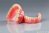
Complete dentures are restorations made for a patient who doesn't have any teeth of their own. After teeth are removed, the bone which was supporting the teeth begins to resorb or shrink away over time. Often this leaves little structure for a denture to rest on, often making the dentures loose and difficult to control.
Complete dentures are typically made out of modern high density plastic materials, including the teeth. Replacement teeth have been developed that are very natural looking and highly esthetic.

The process of making a complete denture is a 4 to 5 step process. At the first appointment, impressions are made using average sized trays. Stone models are made to allow a custom fit tray to be fabricated to use for the second appointment. A very accurate impression is then taken which is used to make the denture. At the third appointment, called the records appointment, measurements are made to allow denture-like appliances made out of plastic and wax to be used to determine the size, shape and color of the teeth along with the correct facial support. At the next appointment, the denture-like appliances are tried in to evaluate the function and esthetics. The teeth have been positioned in wax which allows alterations to be made to the arrangement. Once the doctor and the patient are satisfied with the appearance of the teeth, they are then returned to the dental laboratory for processing. At this time, the wax is exchanged for the pink-colored plastic and the dentures are finished. The dentures are then inserted at the final appointment. During this appointment, the tissue surfaces are carefully evaluated and the teeth are adjusted to ensure the correct bite or occlusion. Additional appointments are scheduled as necessary to adjustments.

Dentures should be evaluated once a year to ensure that the tissues and bone support are healing and functioning properly. Depending upon the patient, dentures typically last several years before needing to either be relined or replaced.
Complete Denture Prosthodontics

Complete Denture Prosthodontics


Selected repeats from Complete Denture Prosthodontics Go through all the questions….. Post your valuable Comments below …… All the best…. Thanks…

Directions:

Click on the correct answer.


Question 1:
For a balanced occlusion, when condylar inclination is increased, the compensating curve should be


Question 2:
In a Complete Denture,the central incisors are generally found to be _______mm anterior to a line bisecting the incisive papilla


Question 3:
In adjusting occlusion for complete denture patient, the two end factors controlling the protrusive movements are


Question 4:
Most commonly used test to check good impression is


Question 5:
By increasing the vertical dimensions of occlusion all with results except


Question 6:
Secondary seal peripherally in lower CD is obtained in area of


Question 7:
While setting condylar guidance on a 3 pin articulator, the incisal pin is


Question 8:
Jiffy denture is a type of


Question 9:
Massetric notch is formed by the action of


Question 10:
As compared to relining, in rebasing of a denture a change is effected in


Question 11:
Squint test is used as guide for selecting


Question 12:
Orientation records are best transferred by


Question 13:
Preservation of what remains is important than meticulous replacement of what is lost quoted by


Question 14:
Stages of polymerization are all except


Question 15:
Boucher's method of relining is a


Question 16:
Which of the following area is not a relief area?


Question 17:
Incomplete flask closure would result in


Question 18:
Most common cause of pain with new dentures is due to


Question 19:
The concept of balanced occlusion is applied primarily to


Question 20:
The main purpose of boxing the impression is to


Question 21:
During polymerization of acrylic resin, above what temperature benzyl peroxide decompose to form free radicals


Question 22:
The intimate contact of the denture periphery with the soft border tissues is referred to as the


Question 23:
The direction of resorption of the maxillary ridge is


Question 24:
During Flasking,_______ mm space should be available between the occlusal surface of the teeth and top of the flask


Question 25:
The labiodental sounds are